BACTERIAL VAGINOSIS
Gallery
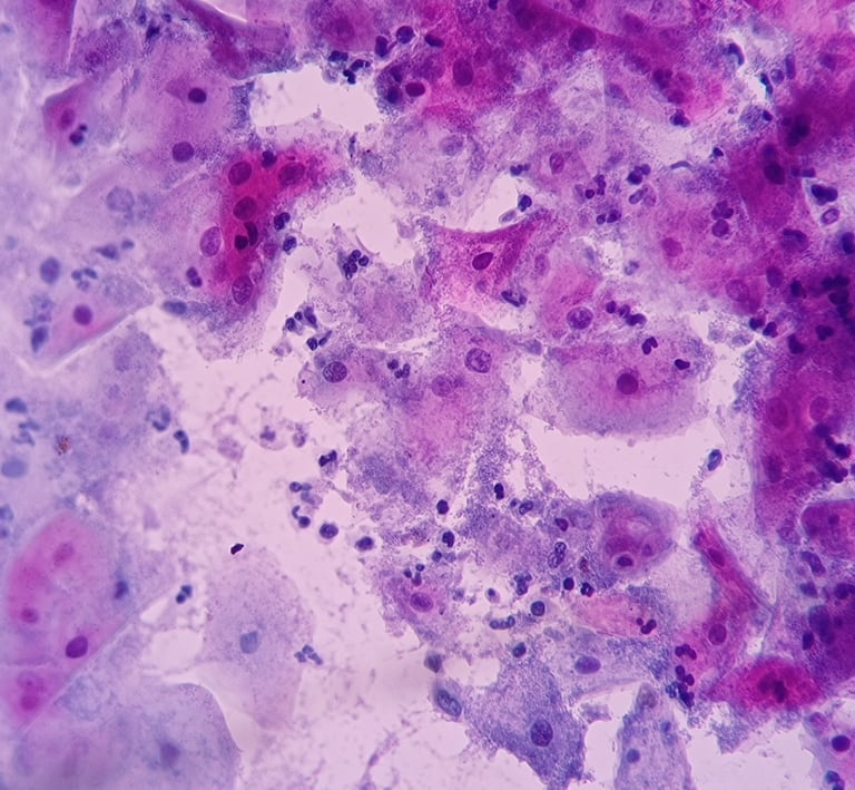
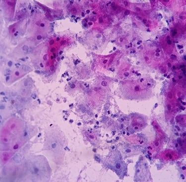
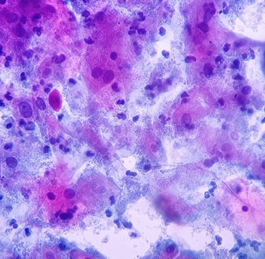
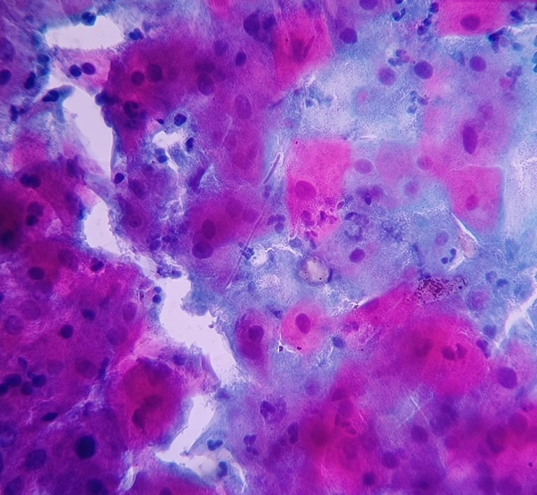
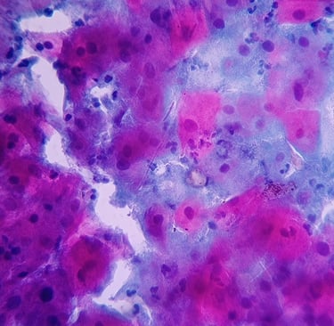
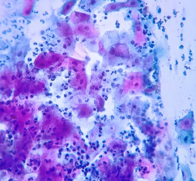
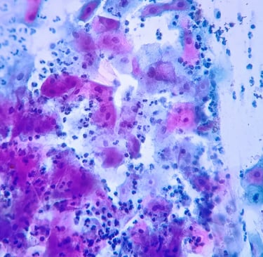
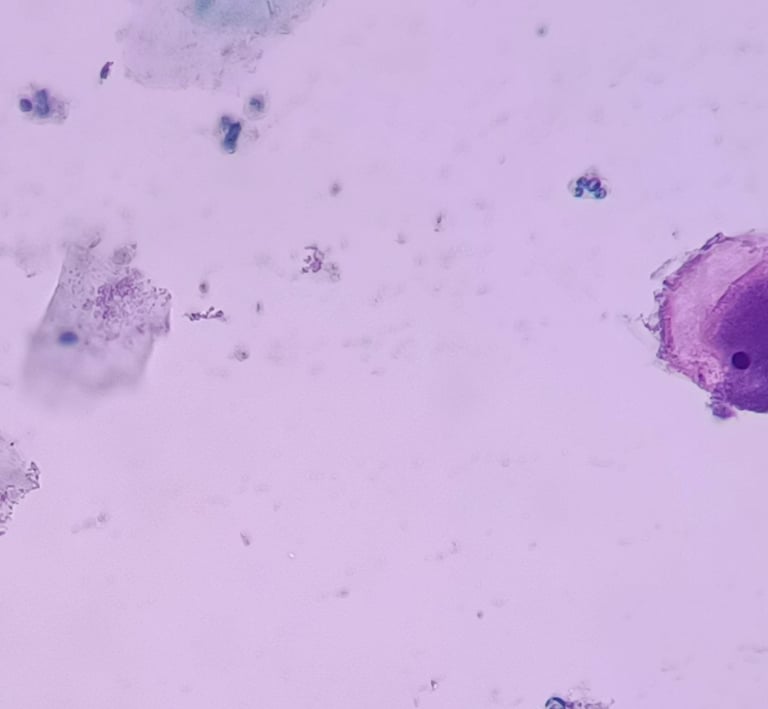
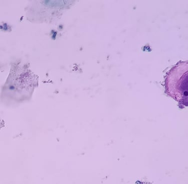
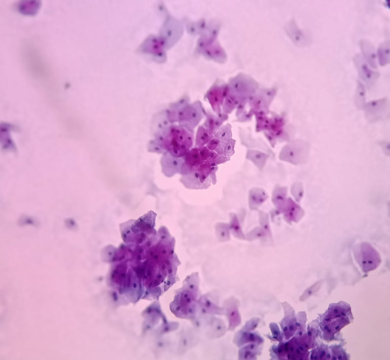
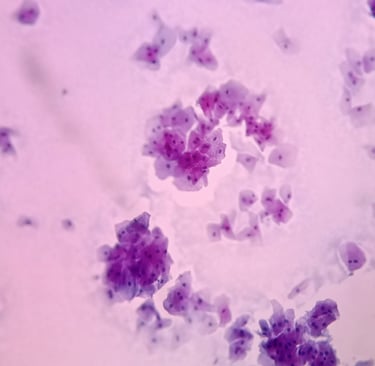
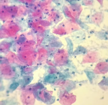
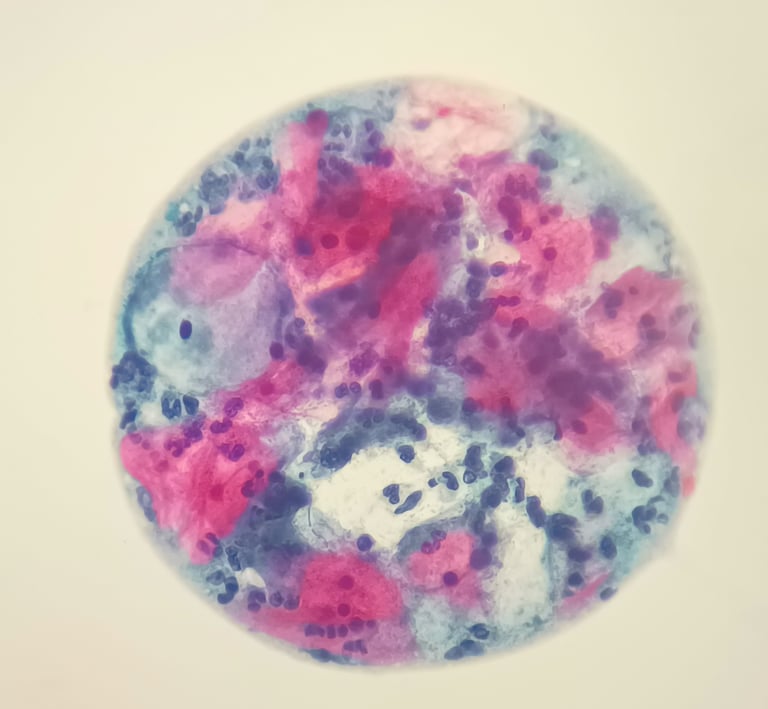
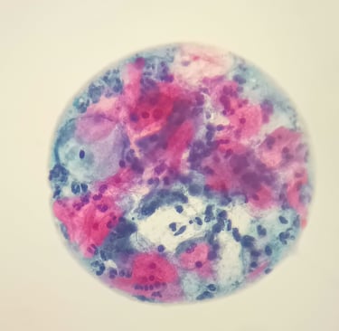
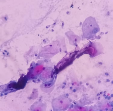
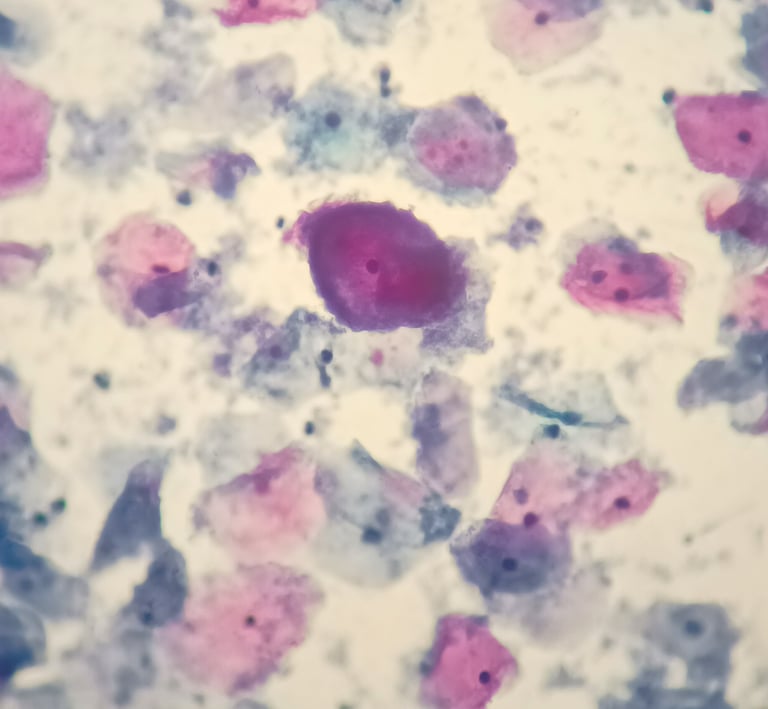
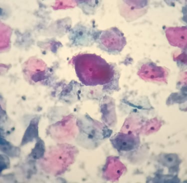
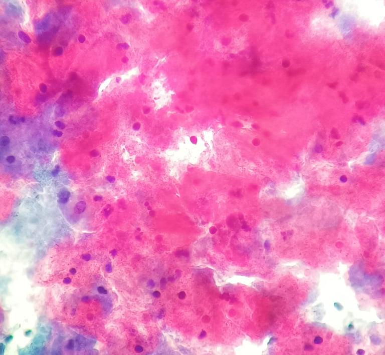
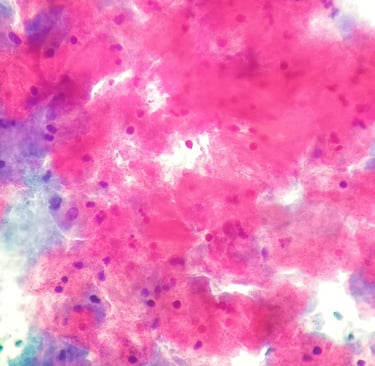
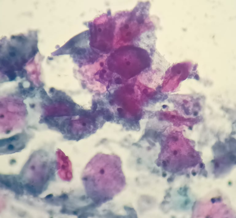
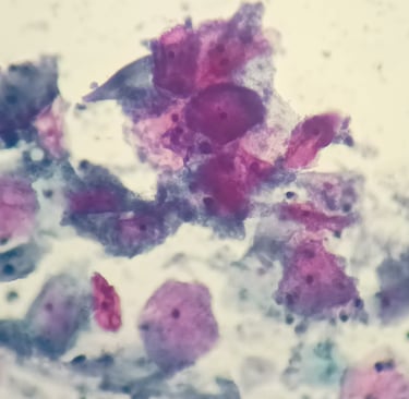
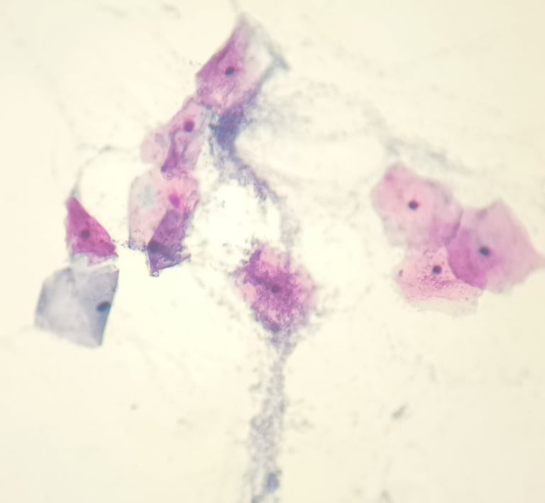
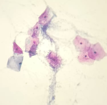
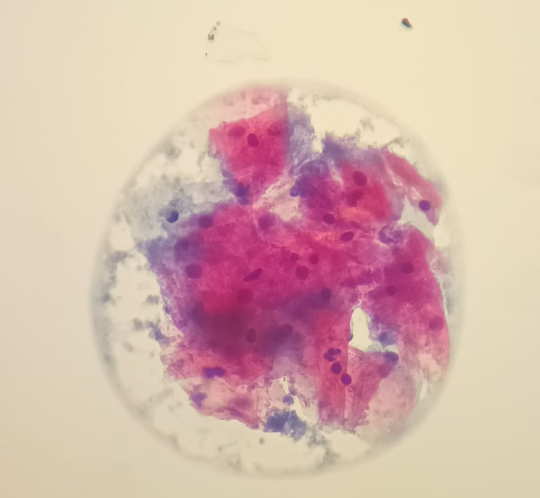
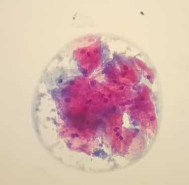
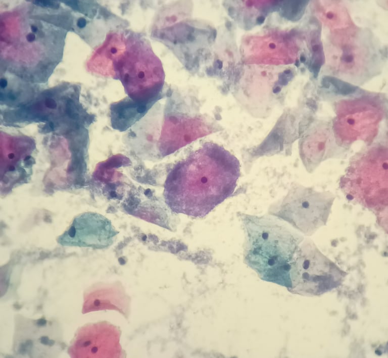
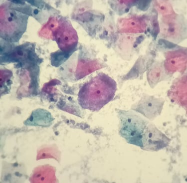
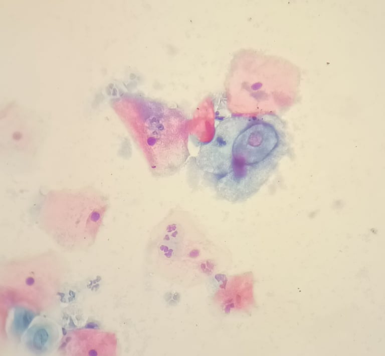
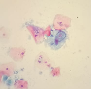
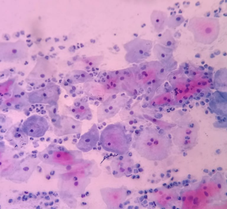
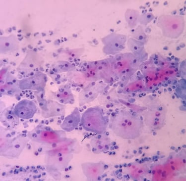
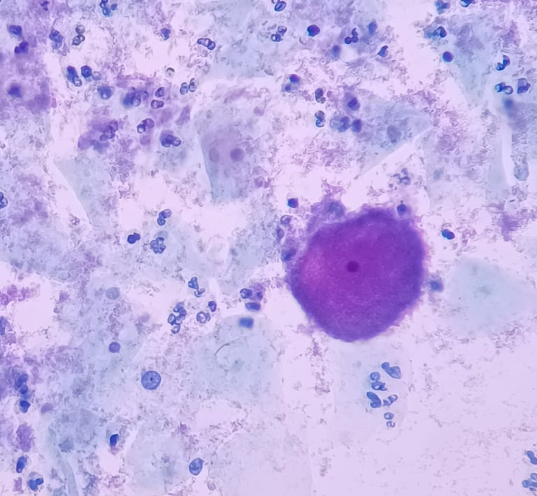
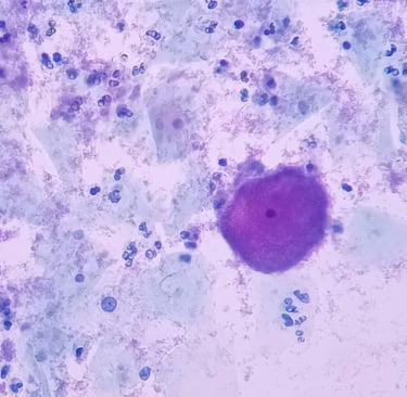
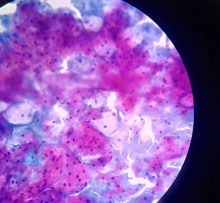
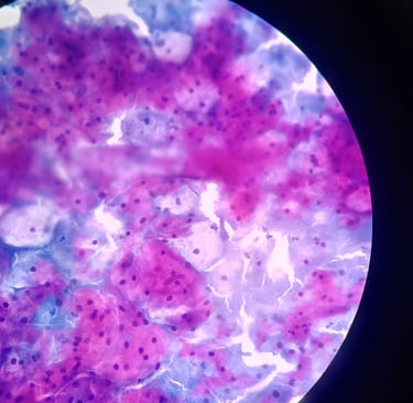
"Individual squamous cells are covered by a layer of coccobacilli that obscure the cell membrane, forming the so-called clue cells. Large numbers of inflammatory cells indicate a vaginitis rather than a vaginosis. There is a conspicuous absence of lactobacilli."
— Nayar, R. & Wilbur, D.C. (Eds.). The Bethesda System for Reporting Cervical Cytology: Definitions, Criteria, and Explanatory Notes. 3rd ed. Springer, 2015.
This website is not affiliated with or endorsed by the authors, editors, or publishers of The Bethesda System for Reporting Cervical Cytology. Content quoted is used strictly for educational and informational purposes.


“The exact cause of BV is not known. While many ‘good’ bacteria are normally found in the healthy vagina, BV results from an imbalance between ‘good’ and ‘harmful’ bacteria micro-organisms. More specifically, an overgrowth of pathogens such as Gardnerella spp., Prevotella spp., Mobiluncus spp., Megasphaera spp., Sneathia spp. and mixed vaginal anaerobes species would eventually replace the ‘beneficial’ lactobacilli that helps maintain a healthy vaginal environment.”
— World Health Organization (2024). Bacterial vaginosis – Fact sheet. Retrieved from https://www.who.int/news-room/fact-sheets/detail/bacterial-vaginosis
This website is not affiliated with or endorsed by the WHO. Quotes are used for educational and informational purposes.
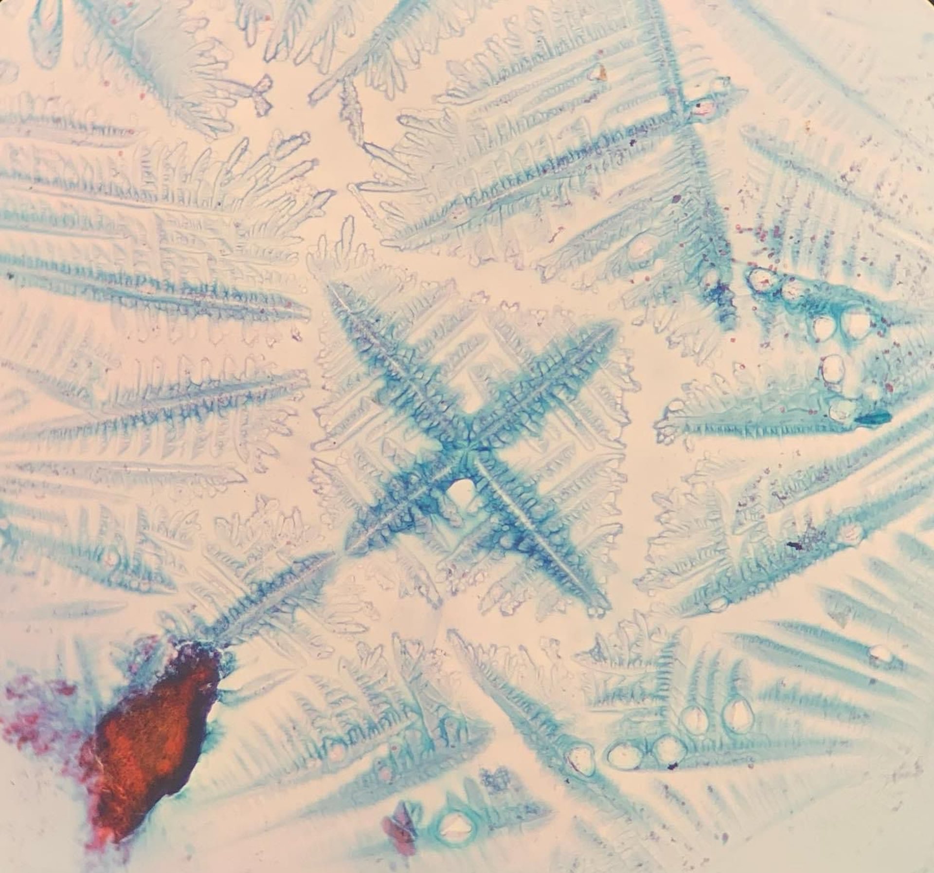
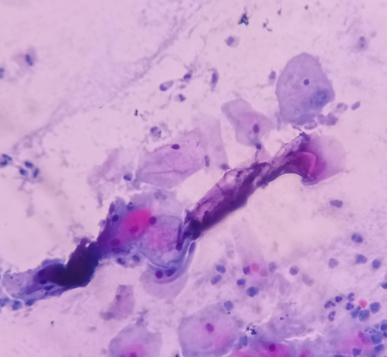
For better interpretation of bacterial vaginosis, it is essential to differentiate between stained mucus, cellular debris from cytolysis, and artifact from Harris hematoxylin residues.
Suggestions or technical problems
Send us your suggestions for improving the site or any technical issues you may have. Thank you...